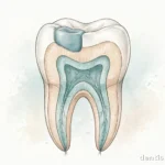
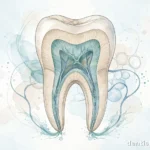
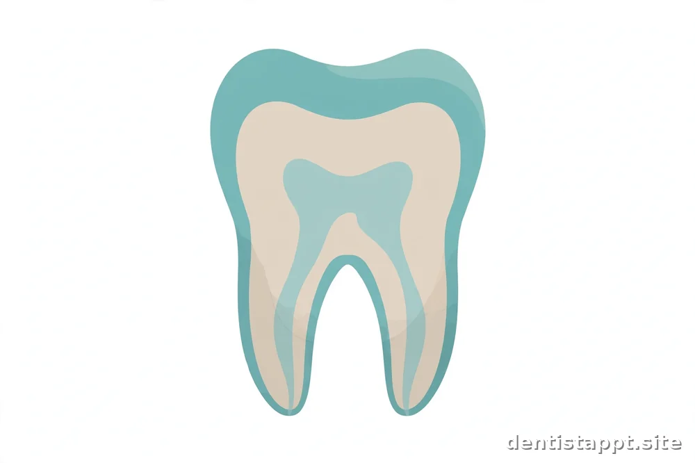
A tooth is made of distinct layers and parts, each with its own job. From the outside in, the three main layers are enamel (the hard outer shell), dentin (the supportive middle layer), and pulp (the living core containing nerves and blood vessels). Understanding these parts gives you the vocabulary to follow conversations about cavities, fillings, and root canals.
The Big Picture: Crown, Neck, and Root
Before diving into layers, it helps to know a tooth's regions. The crown is the visible part above the gumline — the part you brush and chew with. The root is the part anchored below the gumline, held in the jawbone by ligaments. The neck (cervix) is the narrow transition zone where crown meets root, right around the gumline.
A tooth is not a solid block of one material. It is a layered structure, and each layer behaves differently when it is damaged. That is why a small cavity and a deep cavity are treated so differently: they involve different layers, with different properties.
Enamel: The Hard Outer Shell
Enamel is the outermost layer of the crown and the hardest substance in the human body — harder than bone. Its job is protection: it shields the inner layers from the forces of chewing, from temperature changes, and from the acids produced by bacteria and foods.
Two facts about enamel matter for everyday dental care. First, enamel contains no living cells, which means once it is lost, the body cannot regrow it. Acid erosion and decay remove enamel permanently. Second, enamel has no nerves, so decay in enamel alone does not hurt — which is why early cavities are painless and why regular checkups catch problems you cannot feel.
Enamel can be strengthened. Fluoride — from toothpaste, drinking water in many areas, and professional treatments — helps enamel resist acid attack in a process called remineralization. This works on the very earliest stages of mineral loss, before a true cavity forms. Once a cavity has actually opened through the enamel, remineralization can no longer fix it, and a filling is needed. Our guide to dental fillings explains what happens at that stage.
Dentin: The Supportive Middle Layer
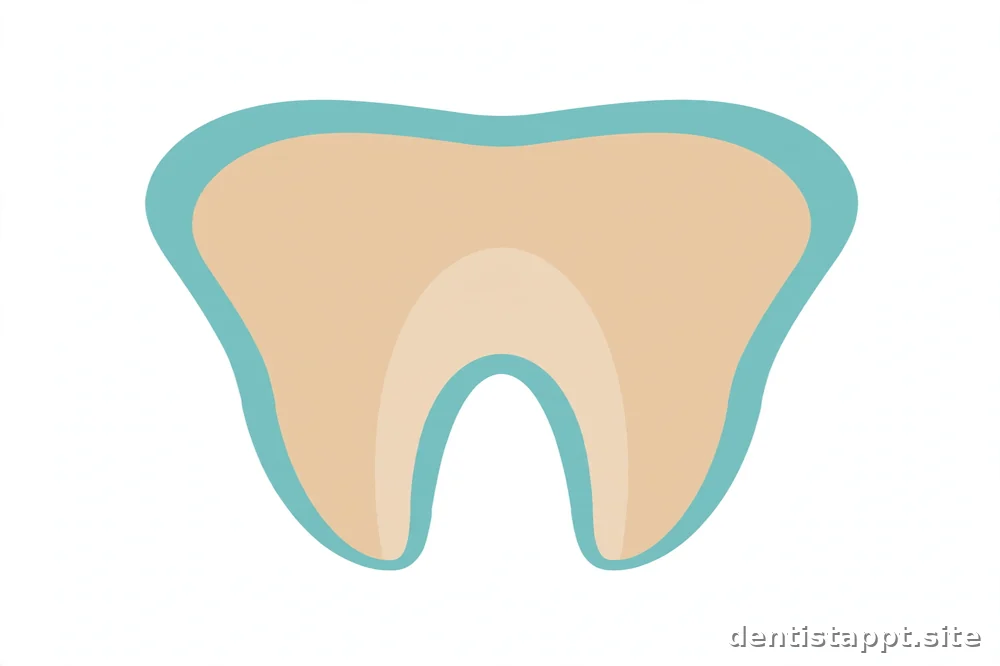
Beneath the enamel lies dentin, a softer, yellowish layer that forms the bulk of the tooth's structure. Dentin is less mineralized than enamel, which makes it somewhat flexible — it absorbs chewing forces and supports the rigid enamel shell, a bit like the foam core under a hard outer panel.
Unlike enamel, dentin is alive in a limited sense: it contains microscopic tubules (tiny channels) that run from the enamel inward to the pulp. These tubules are why decay accelerates once it reaches dentin — bacteria travel through the tubules more easily than through solid enamel. The tubules are also why deep cavities hurt: stimuli such as cold, heat, or sweets reach closer to the nerve through these channels.
Dentin forms continuously throughout life. When a tooth is irritated — by decay, wear, or repeated dental work — it can lay down extra "reparative" dentin as a defensive response, thickening the barrier between the irritation and the pulp. This is one reason dentists monitor worn or cracked teeth over time rather than treating every sign of wear immediately.
Pulp: The Living Core
At the center of the tooth is the pulp: soft tissue containing nerves, blood vessels, and connective tissue. The pulp occupies the pulp chamber in the crown and extends down through root canals in the root. In a young tooth, the pulp is large; as the tooth matures and lays down more dentin, the pulp space gradually shrinks.
The pulp's roles include nourishing the tooth during development and providing sensation — the reason a tooth can feel hot, cold, and pain. In a fully developed adult tooth, the pulp's ongoing role is smaller, but it still matters: a healthy pulp helps the tooth respond to injury and maintain its internal defenses.
When decay or trauma reaches the pulp, infection can develop inside the tooth. Because the pulp sits in a rigid chamber with no room to swell, pressure builds and pain can become severe. This is the stage at which root canal treatment enters the picture: the procedure removes the infected pulp, cleans and seals the canal space, and saves the tooth's structure. Our concept-level guide to root canal treatment explains the idea in plain terms.
Cementum: The Root's Covering
Cementum is a thin, bone-like layer covering the root, where enamel does not reach. It is softer than enamel and its main job is to give the periodontal ligament — the fibers that anchor the tooth to the jawbone — something to attach to.
Cementum matters in daily life when gums recede. Gum recession can expose the root surface, and exposed cementum wears and decays more easily than enamel. That is one reason dentists pay attention to gum health and brushing technique: aggressive brushing can contribute to recession and expose this vulnerable layer.
The Supporting Cast: Beyond the Tooth Itself
A tooth does not stand alone. Several structures hold it in place and keep it functional:
- Periodontal ligament — a sling of fibers connecting the cementum to the jawbone. It acts as a shock absorber during chewing and gives teeth their slight natural movement.
- Alveolar bone — the jawbone socket that holds the root. Bone responds to pressure, which is how orthodontic tooth movement works, but bone loss from gum disease loosens teeth.
- Gums (gingiva) — the soft tissue sealing around the neck of each tooth, protecting the underlying bone and ligament from bacteria.
- Nerves and blood supply — entering through the tip of the root, serving the pulp.
Dental health means the health of this whole system, not just the visible crown. Gum disease, for example, attacks the supporting structures while the tooth itself may look fine — which is why dentists measure gum pockets and check bone levels, not just the teeth.
How the Parts Work Together: A Chew-by-Chew View
Consider what happens when you bite into an apple. The enamel takes the initial force and resists the fruit's acidity. The dentin underneath flexes slightly, absorbing shock so the rigid enamel does not crack. The periodontal ligament lets the tooth shift a fraction of a millimeter, distributing the load to the alveolar bone. The pulp registers the cold and the pressure, giving your brain feedback. The gums seal the whole assembly against bacteria in the mouth.
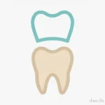
Every common dental problem can be mapped onto these parts. A cavity is decay progressing through enamel into dentin (and potentially pulp). A filling replaces lost enamel and dentin with restorative material. A crown caps a tooth whose enamel and dentin can no longer support chewing on their own. Gum disease is an attack on the supporting cast. This vocabulary is the foundation for understanding nearly every other dental topic.
Why This Anatomy Matters for Patients
You do not need to memorize tooth anatomy, but a basic grasp of it changes how you hear a dentist's explanations. "The decay is still in the enamel" means a small, likely simple fix. "It has reached the dentin" means the cavity is progressing and needs attention. "The pulp is involved" means the treatment conversation changes significantly. Knowing the layers helps you ask better questions and understand why the dentist recommends what they recommend.
It also explains prevention. Brushing with fluoride toothpaste protects enamel. Flossing protects the contact areas where enamel is thin and decay starts easily. Regular checkups catch enamel-stage problems before they reach dentin. Every preventive habit maps to protecting a specific part of this structure.
FAQ
Which part of the tooth has nerves?
The pulp, the soft tissue at the center of the tooth, contains the nerves (along with blood vessels). Enamel has no nerves at all, which is why early decay does not hurt. Dentin has no nerves either, but its microscopic tubules transmit stimuli toward the pulp, which is why deep cavities or exposed dentin can cause sensitivity to hot, cold, or sweet foods.
Can enamel grow back once it is lost?
No. Enamel contains no living cells, so the body cannot regenerate it. Fluoride can help remineralize enamel in the very earliest stage of mineral loss, before a cavity forms, but once enamel is actually broken through or worn away, it is gone permanently. This is why dentists emphasize preventing enamel loss through brushing, fluoride, and limiting acidic and sugary foods.
What is the difference between dentin and pulp?
Dentin is the hard, supportive middle layer of the tooth — mineralized tissue with microscopic tubules running through it. Pulp is the soft living tissue at the very center, containing nerves and blood vessels. Dentin surrounds and protects the pulp. Decay moves through dentin faster than through enamel because of the tubules, and once it reaches the pulp, infection and significant pain can follow.
What holds a tooth in the jawbone?
The root is held in its socket (the alveolar bone) by the periodontal ligament, a network of fibers attaching the root's cementum covering to the bone. The ligament acts as a shock absorber during chewing. Gum disease can destroy the ligament and bone, which is why advanced gum disease causes teeth to loosen even when the teeth themselves are intact.
Do baby teeth have the same parts as adult teeth?
Yes, the basic structure is the same: enamel, dentin, pulp, cementum, and supporting structures. The main differences are proportion — baby teeth have thinner enamel and relatively larger pulp chambers, which means decay can reach the pulp faster. Their roots are also designed to dissolve (resorb) as the permanent teeth develop underneath. The thinner enamel is one reason dentists watch children's teeth closely.
This site is educational information only — not medical advice. Consult a qualified dental professional about your own situation.